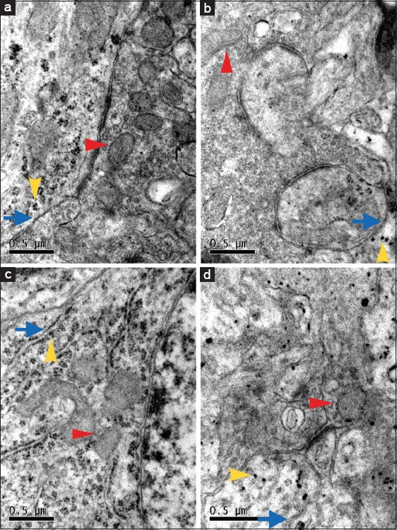
Figure 3.

The ultrastructure of neurons in hippocampal CA1 region under electron microscope in sham operated C3H/HeN mice (a) and cardiopulmonary resuscitation treated C3H/HeN mice (b) and sham operated C3H/HeJ mice (c) and cardiopulmonary resuscitation treated C3H/HeJ mice (d). The red arrow indicates mitochondria, the yellow arrow indicates ribosome and the blue arrow indicates endoplasmic reticulum. The mitochondria and endoplasmic reticulum appeared more swollen with the reduction of ribosome in b and d (n = 4, per group). Scale bar = 0.5 μm.
