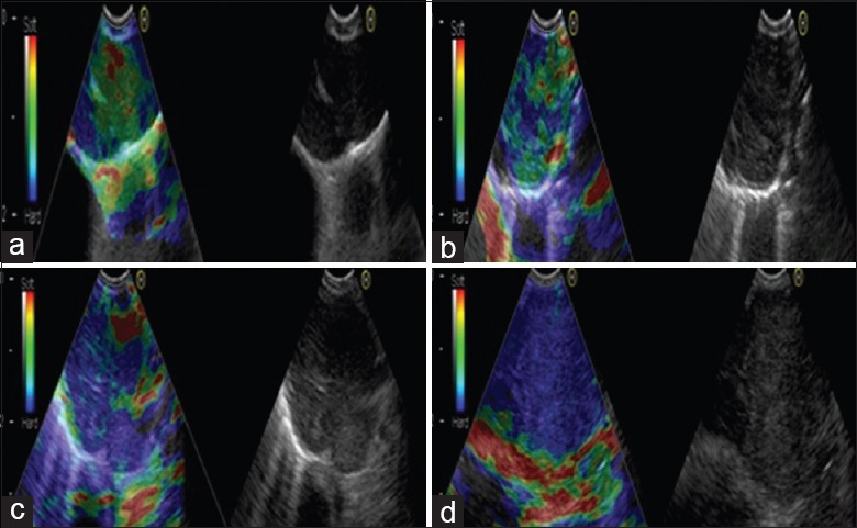
Figure 1.

Representative lymph nodes on endobronchial ultrasound elastography. (a) Representative images showing that the lymph node had a distinct boundary, low echo, and homogeneous echo. The elastography grading score in this figure was 1 point. Histopathological specimen from endobronchial ultrasound-guided transbronchial needle aspiration demonstrated the existence of inflammation. (b) Representative images showing that the lymph node was round, the boundary was clear and the internal echo was low. The herein elastography grading score was 2 points. The pathological result confirmed the existence of granuloma lesion. (c) Representative images showing that the lymph node had a distinct boundary, medium echo and uneven echo. The elastography grading score in this figure was 3 points. The pathological result showed the diagnosis of small cell lung cancer. (d) Representative images showing that the lymph node had a distinct boundary, medium echo and uneven echo. The herein elastography grading score was 4 points. The pathological result showed the diagnosis of poorly differentiated adenocarcinoma.
