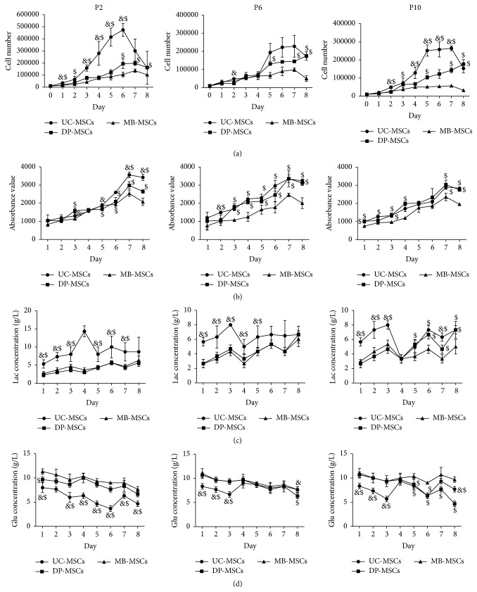
Figure 3.
Growth kinetics of UC-, DP-, and MB-MSCs. (a) The cell number determined on an automatic cell counter; (b) cell viability determined by Alamar Blue staining; (c-d) the glucose (Glu) and lactate (Lac) levels monitored using the glucose and lactate biosensors. Each experiment was performed in triplicate: &compared with DP-MSCs at corresponding day; $compared with MB-MSCs at corresponding day, p < 0.05. UC-MSCs: umbilical cord mesenchymal stem cells; DP-MSCs: dental pulp mesenchymal stem cells; MB-MSCs: menstrual blood mesenchymal stem cells; P: passage.

