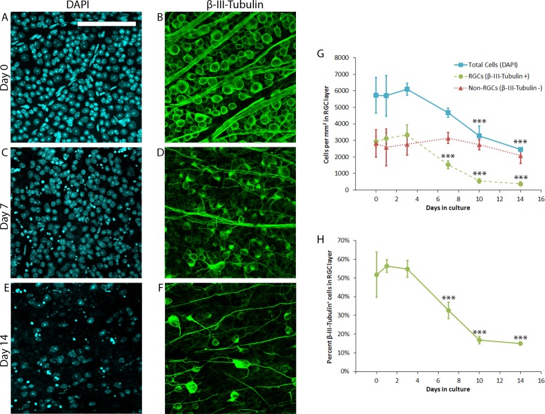
Figure 1.
Characterization of RGC loss in mouse retinal explants. Wild-type CD1 mouse retinal explants (N = 5 explants per time point) were cultured for 0, 1, 3, 7, 10, or 14 days prior to fixation and immunofluorescent labeling of all cells (DAPI+, [A, C, E]) or RGCs (β-III-Tubulin+, [B, D, F]). Representative micrographs from nonoverlapping fields are shown in (A–F); scale bars: 100 μm. Quantification of DAPI+ and β-III-Tubulin+ cell density is presented in G. Non-RGCs represent the difference between DAPI+ and β-III-Tubulin+ cells. Percent β-III-Tubulin+ cells in the RGC layer is presented in (H). Error bars represent SD. ***P ≤ 0.001 by Dunnett's post hoc test compared with day 0 time point.

