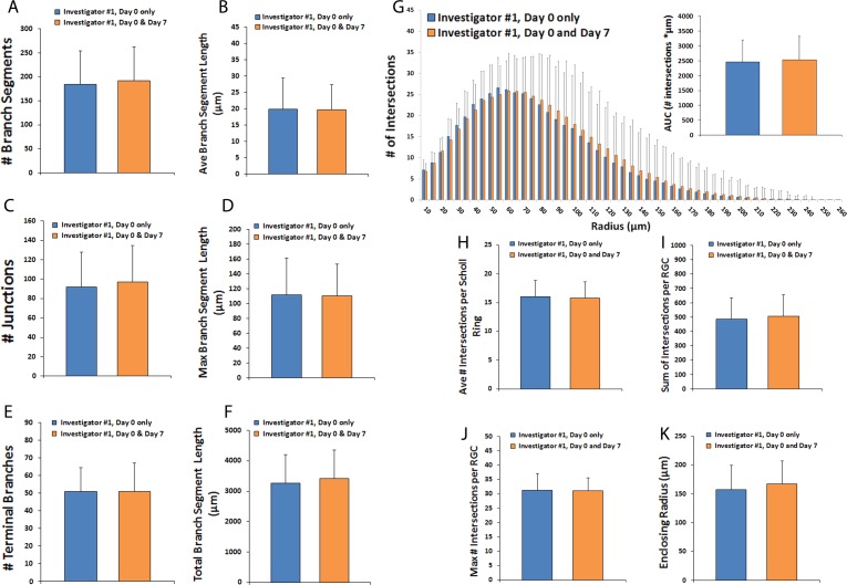
Figure 4.
Comparison of dendritic fields in RGCs living versus dead after 7 days in culture. Thy1-YFP retinal explants were cultured for 7 days. Z-stack confocal images of live YFP+ RGCs (N = 130 RGCs) were obtained on day 0 only and cells were divided according to whether they were still alive and visible by microscopy at 7 days (N = 75 RGCs) or were no longer detectable at 7 days (N = 55 RGCs). Dendritic field parameters (A–F) for RGCs imaged at day 0 are shown. Scholl histograms (G) depict the number of intersections between dendritic fields and Sholl rings of increasing eccentricity; inset graph quantifies the AUC of the Scholl histogram. Sholl parameters for day 0 images are shown (H–K). Error bars represent SD. P > 0.05 by unpaired t-test for all comparisons.

