Figure 6.
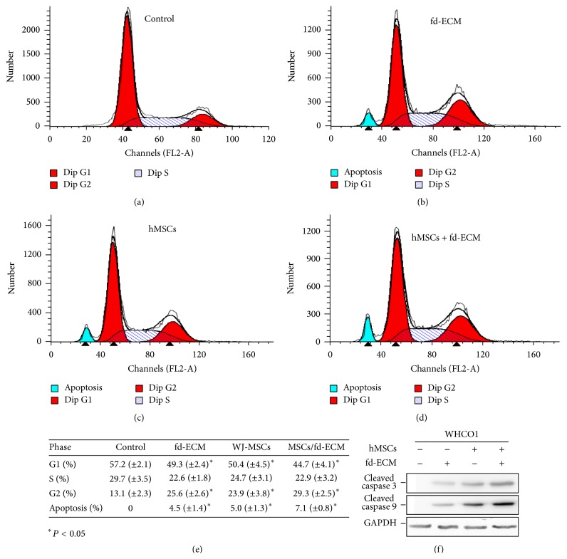
MSCs and fd-ECM synergistically induce cancer cell apoptosis in vitro. (a) Flow cytometric analysis of control WHCO1 cells showed no apparent apoptosis after 48 hrs of incubation. (b) WHCO1 cells were cultured on fd-ECM for 48 hrs and harvested and stained with propidium iodide for cell cycle analysis using flow cytometry. (c) WHCO1 cells were cocultured with MSCs for 48 hrs, harvested, and stained with propidium iodide for cell cycle analysis using flow cytometry. (d) Cocultured WHCO1 cells were plated on an fd-ECM for 48 hrs, harvested, and stained with propidium iodide for cell cycle analysis using flow cytometry. (e) Percentage of cells in each stage of the cell cycle after WHCO1 cells were cultured in MSC-CM, cocultured with MSCs, and cultured on fd-ECM. Four different experiments were pooled together. Data are presented as mean ± standard deviation. ∗ P < 0.05. (f) MSCs and fd-ECM synergistically induce cleaved caspases 3 and 9 expression in WHCO1 cells.

