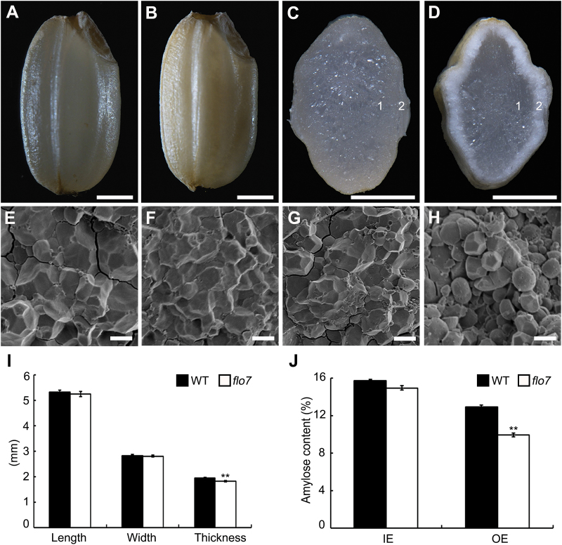
Fig. 1.
Phenotypic analyses of the flo7 mutant. (A–D) Appearance and transverse sections of representative wild-type (A, C) and flo7 mutant (B, D) dry seeds. Bars, 1mm. (E–H) Scanning electron microscopy images of transverse section of the wild-type (E, F) and flo7 mutant (G, H) grains. (E) and (F) represent the magnified region indicated by ‘1’ and ‘2’ in the wild-type endosperm, respectively, and (G) and (H) represent the magnified region marked by ‘1’ and ‘2’ in the flo7 mutant endosperm, respectively. Bars, 5 μm. (I) Measurement of seed length, seed width, and seed thickness of wild-type and flo7 mutant grains (n=20 each). (J) Amylose content comparison of the wild-type and flo7 mutant endosperm parts (n=3 each). IE, inner endosperm, OE, outer endosperm. Data are given as means±SD (from at least three independent samples) and were compared with wild type by Student’s t-test (**P<0.01).

