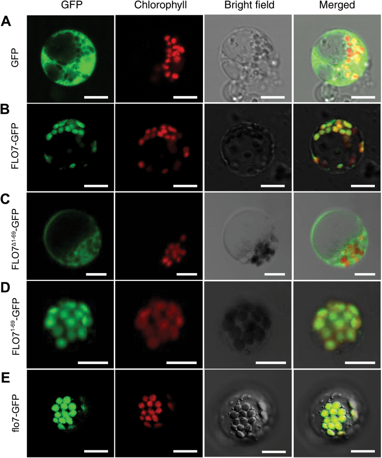
Fig. 6.
) Confocal microscopy images showing the subcellular localization of FLO7 in rice protoplasts. (A) GFP by itself localized to the cytoplasm and nucleus. (B) Full-length FLO7 fusion protein (FLO7–GFP, aa 1–364) localized to the chloroplasts. (C) FLO7 fusion protein lacking the N-terminal 69 aa (FLO7Δ1–69–GFP; aa 70–364) displayed a diffuse localization pattern. (D) The N-terminal 69 aa of FLO7 (FLO71–69–GFP) were capable to target GFP proteins to the chloroplasts. (E) Mutation of the FLO7 protein in the flo7 mutant (flo7–GFP, aa 1–336) failed to impair its chloroplast-localization pattern. After 16h of incubation, rice protoplasts were observed using a confocal laser scanning microscope. GFP (green), chlorophyll autofluorescence (red), bright-field images, and an overlay of green and red signals are shown. Bars, 10 μm.

