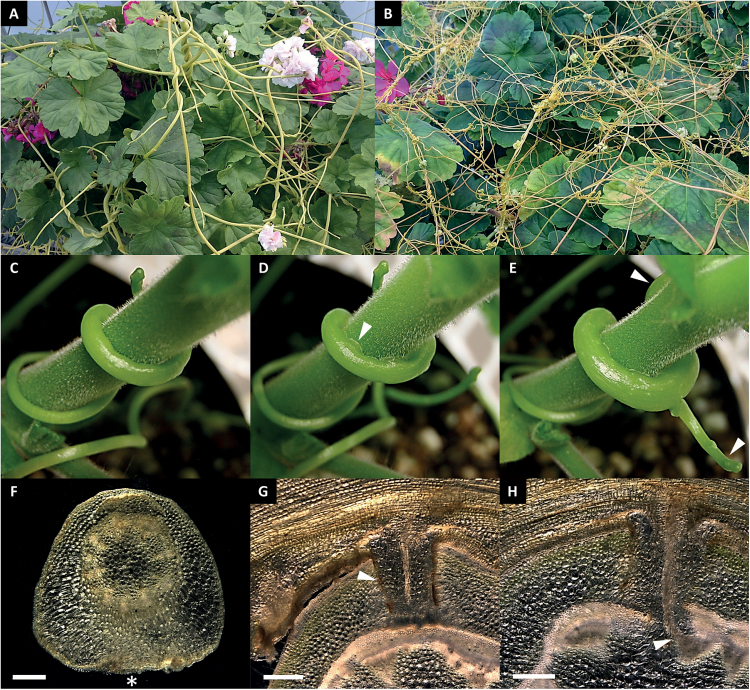
Fig. 1.
Cuscuta infecting the compatible host plant P. zonale. (A) C. reflexa and (B) C. gronovii parasitizing P. zonale. The infection of P. zonale by C. reflexa can be divided into three stages. (C and F) In the first swelling stage the parasite stem grows in width at the side facing the host (asterisk). (D and G) After attachment to the host surface (arrowhead in D), the haustorium (arrowhead in G) invades the host tissue (penetrating stage). (E and H) The mature stage is defined by established connections between the vascular systems of the two plants (arrowhead in H) and is recognized by apical shoot growth and the formation of additional side shoots (arrowheads in E). Cross-sections were made with respect to orientation of the parasite shoot axis in (F), and with respect to the host shoot axis in (G) and (H). Scale bars are 500 µm.

