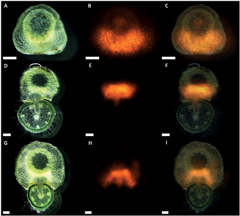
Fig. 9.
XET action during infection of P. zonale by C. reflexa. Cross-sections of (A) swelling, (D) penetrating, and (G) mature infection stages tissue printed on XET test paper. (B, E, and H) Fluorescence micrographs showing XET action in the printed tissues in (A), (D), and (G), respectively. Merged pictures of (A–B), (D–E), and (G–H) are presented in (C), (F), and (I), respectively. Scale bars are 500 µm. Visible fibres are integral to test papers.

