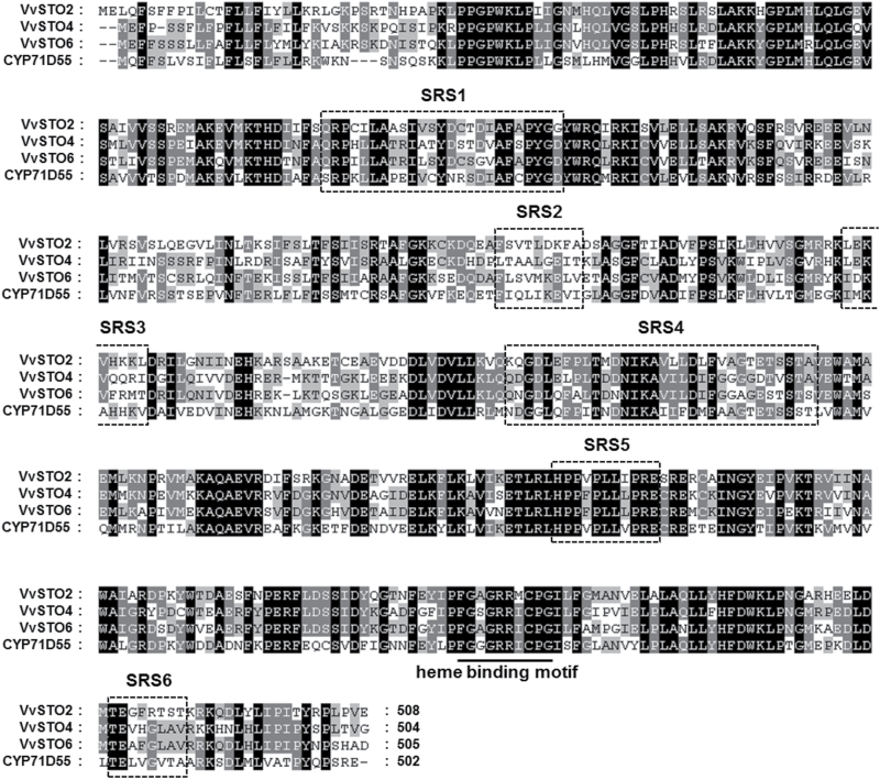
Fig. 1.
Amino acid sequence alignment of VvSTO2, VvSTO4, VvSTO6 and CYP71D55. Multiple sequence alignment was performed using ClustalW and visualized by the software Genedoc. Six of the putative SRS regions defined by Gotoh (Gotoh, 1992) are outlined and the heme-binding motifs are underlined and annotated. The black and gray shades indicate similar amino acids, respectively.

