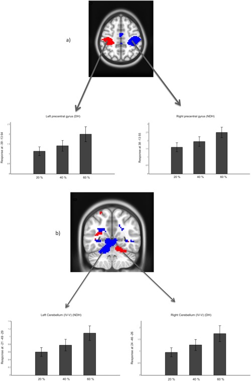
Figure 5.

One sample t tests at the group level of Linear effect responses using DH (red) and NDH (blue) in (a) precentral gyri; (b) cerebellum IV–V. All clusters are corrected for multiple comparisons after using a threshold of 0.001 (uncorrected) at the voxel level. In the images, right is right and left is left; axial cut at z = 50; coronal cut at y = −54. [Color figure can be viewed in the online issue, which is available at http://wileyonlinelibrary.com.]
