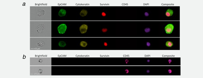
Figure 1.

EpCAM, cytokeratin, survivin and CD45 expression in oesophageal adenocarcinoma cells and in white blood cells. SK‐GT‐4 cells were grown to 80% confluence in routine culture medium, trypsinised and 1 × 10⁶ cells fixed with 1% formalin. Cells were permeabilised by incubation with 0.3% saponin, incubated overnight with antibodies against cytokeratins 4, 5, 6, 8, 10, 13 and 18, and CD45, and incubated subsequently with antibodies against EpCAM and survivin. Cells were washed, re‐suspended in 100 μl and 2 μl DAPI added. Cells were visualised with an ImageStreamX flow cytometer with the lasers set to emit excitation at 405, 488, 561 and 658 nm (a). Red blood cells were removed from whole blood by ammonium chloride lysis, the remaining blood cells were concentrated by centrifugation, fixed, permeabilised and incubated with antibodies against EpCAM, cytokeratins 4, 5, 6, 8, 10, 13 and 18, survivin and CD45 as in (a). Blood cells were concentrated, incubated with DAPI and visualised (b). Images were collected with a ×40 objective with the wavelengths for the collection channels set at: 480–560 nm, EpCAM; 560–595 nm, cytokeratins; 745–800 nm, CD45; 430–505 nm, DAPI; and 642–745 nm, survivin.
