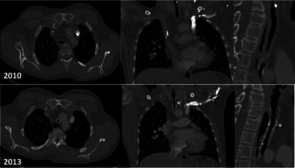
Figure 5.

Computer tomography images: axial, coronal, and sagittal sections taken in 2010 and 2013. The stents can be seen embedded within the tracheal wall in the 2013 scans (*). The narrowed transplanted segment is visible in both the 2010 and 2013 images. The left sided superior vena cava is not unusual in patients with long segment congenital tracheal stenosis.
