Figure 2.
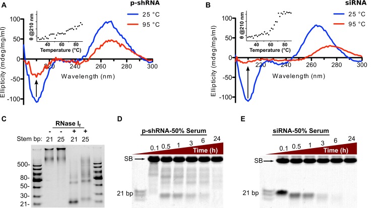
Characterizing the secondary structure and stability of p-shRNA. (A and B) CD spectra of p-shRNA (21 bp stem/10 base loops) and siRNA (21 bp with same sequence) measured at 25 and 95°C. The insets plot the CD signal at 210 nm as a function of temperature. (C) p-shRNAs with 21 or 25 bp stems (from templates 6 and 12, respectively) were treated with RNase I for 15 min then run on a native 15% TBE-PAGE gel. The observed banding pattern confirms the predicted alternating single-/double-stranded structure of p-shRNA. (D and E) p-shRNA (21 bp stem/10 base loops; from template 6) and siRNA (21 bp) were treated with 50% human serum for 0.1–24 h and run on a native 15% TBE-PAGE gel (SB indicates background serum band). Ladders = dsRNA ladder (NEB) and siRNA marker (NEB).

