Figure 6.
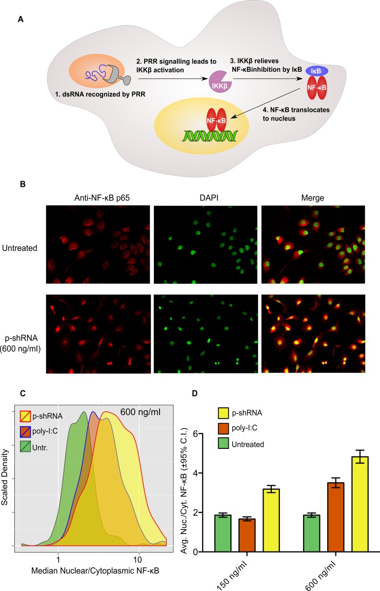
Activation of NF-κB in SKOV3 cells by p-shRNA-25. (A) NF-κB is activated by dsRNA through pattern recognition receptor (PRR)-mediated signaling, resulting in nuclear translocation and transcriptional activation of myriad genes. (B) Fluorescence imaging of SKOV3 cells stained with anti-NF-κB (red) and DAPI (green). Poly-I:C and p-shRNA were complexed with Lipofectamine and incubated with cells at the indicated concentrations for 1 h prior to fixation and staining. The yellow color in the merged images indicates nuclear localization of NF-κB. (C and D) Nuclear localization of NF-κB was quantified using Cell Profiler by taking the ratio of median NF-κB fluorescence in the nucleus divided by median NF-κB fluorescence in the cytoplasm for untreated cells or cells treated with 150 or 600 ng/ml RNA/Lipofectamine complexes (for histograms of samples treated with 150 ng/ml see Supplementary Figure S11). Values in (D) correspond to the geometric means of the histograms ±95% confidence intervals.

