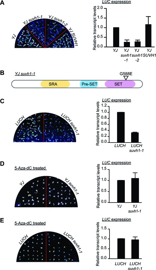
Figure 1.

Characterization of suvh1 mutants. (A) The suvh1–1 mutation led to decreased expression of the luciferase gene (LUC) in the YJ background. (Left panel) LUC luminescence of 8-day-old YJ, YJ suvh1–1, YJ suvh1–2 and YJ SUVH1 seedlings grown on MS media. (Right panel) RT-qPCR revealed decreased LUC transcript levels in the suvh1 mutants in the YJ background. ‘YJ SUVH1’ indicates the YJ SUVH1p:SUVH1–3XFLAG suvh1–1 line. In YJ SUVH1, the phenotype of the YJ suvh1–1 mutant was rescued by the transgene containing a wild-type SUVH1 genomic region and a 3XFLAG tag at the C-terminus of SUVH1. (B) A diagram of the SUVH1 protein and the substitution caused by the suvh1–1 mutation. The SUVH1 protein contains an SRA domain, a Pre-SET domain and a SET domain. The G-to-E substitution caused by suvh1–1 occurs in the SET domain. (C) The suvh1–1 mutation led to decreased LUC expression in the LUCH background. (Left panel) LUC luminescence of 8-day-old LUCH and LUCH suvh1–1 seedlings grown on half-strength MS media. (Right panel) RT-qPCR showed decreased LUC expression in the suvh1–1 mutant in the LUCH background. (D–E) The effects of suvh1–1 on LUC expression were suppressed by 5-Aza-2′-deoxycidine treatment in both the YJ (D) and LUCH (E) backgrounds. (Left panels) LUC luminescence of seedlings grown on half-strength MS media containing 7 μg/ml 5-Aza-2′-deoxycytidine for 14 days for YJ and YJ suvh1–1 (D) and LUCH and LUCH suvh1–1 (E). (Right panels) RT-qPCR showed rescued LUC transcript levels in the treated seedlings. Error bars in (A–E) represent standard deviation from three biological replicates.
