
Figure 3.
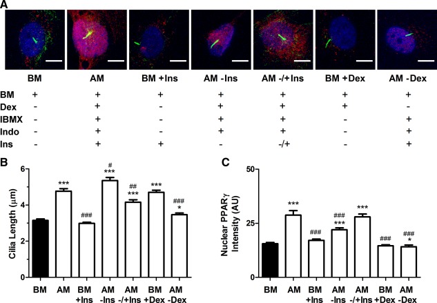
The role of media components in cilia elongation and adipogenic differentiation. (A): Representative confocal images showing primary cilia and nuclear peroxisome proliferator‐activated receptor γ (PPARγ) expression in human mesenchymal stem cells (hMSCs) cultured for 2 days in the presence of different media components: basal media (BM), complete adipogenic media (AM) including insulin, BM with insulin (BM+Ins), adipogenic media without insulin (AM − Ins), adipogenic media with no insulin for the initial 24 hours followed by addition of insulin for 24 hours (AM ± Ins), BM with dexamethasone alone (BM+Dex) and adipogenic media without dexamethasone (AM‐Dex). PPARγ is shown in red, acetylated α‐tubulin in green, and Hoechst stained nuclei in blue. Scale bar = 10 µm. Cilia length (B) and nuclear PPARγ intensity (C). n = 100–110 cilia per group in (B) and n = 100–110 nuclei per group in (C). *, p < 0.05 versus BM; ***, p < 0.001 versus BM; #, p < 0.05 versus AM; ##, p < 0.01 versus AM; ### p < 0.001 versus AM; Mann–Whitney U test. Abbreviations: AM, adipogenic media; BM, basal media; Dex, dexamethasone; IBMX, 3‐Isobutyl‐1‐methylxanthine; Ins, insulin; PPARγ, peroxisome proliferator‐activated receptor γ.