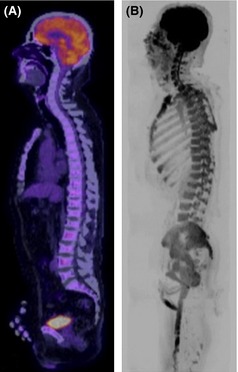
Figure 1.

Diffusion weighted magnetic resonance imaging (DW‐MRI) shows increased sensitivity for diffuse marrow infiltration. Sagittal 18F‐fluorodeoxyglucose positron emission tomography (18F‐FDG PET)/computerized tomography (CT) (A) and b900 whole body DW‐MRI Maximum Intensity Projection image in a 52‐year‐old male with multiple myeloma. (A) 18F‐FDG PET/CT was reported as normal with no areas of increased FDG uptake and no lytic lesions on the CT component. (B) Inverted greyscale WB DW‐MRI demonstrated diffuse marrow infiltration. Marrow trephine confirmed 80–90% infiltration with plasma cells.
