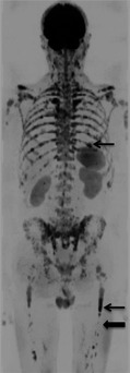
Figure 2.

Whole body diffusion weighted magnetic resonance imaging (WB DW‐MRI) provides excellent contrast between normal bone marrow and focal lesions. b900 WB DW‐MRI Maximum Intensity Projection image in a 55‐year‐old male with multiple myeloma demonstrates multiple focal lesions that appear low signal on the inverted grey scale image (examples indicated by arrows) with excellent contrast against normal marrow, which does not return signal (block arrow).
