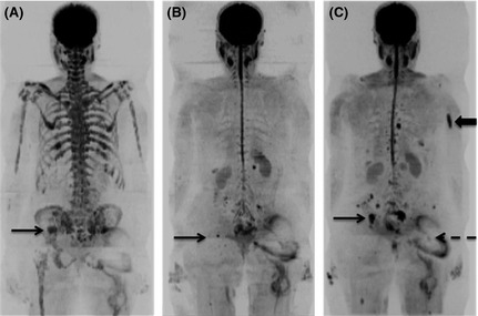
Figure 5.

The extremely high sensitivity of diffusion weighted magnetic resonance imaging (DW‐MRI) allows detection of tiny foci of residual disease post‐autograft. b900 whole body DW‐MRI Maximum Intensity Projection images of a 70‐year‐old female with multiple myeloma before (A), 3 months post‐ (B) and 8 months post‐ (C) autograft. At baseline there is widespread marrow disease including a focal lesion in the right iliac bone (A, arrow). At 3 months post‐autograft, the marrow signal has normalized apart from a tiny residual focus of abnormal signal at a site of original disease in the right iliac bone (B, arrow). WB DW‐MRI 8 months post‐autograft shows progression of the residual disease (C, arrow) and development of new sites (example shown by block arrow). Ill‐defined distortion overlying the left hip (dashed arrow) represents artefact from a metal hip prosthesis.
