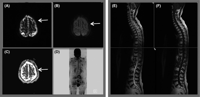
Figure 6.

Whole body MRI protocol. Diffusion weighted magnetic resonance imaging (DW‐MRI) is performed axially using b values of 50 (A) and 900 (B). The system software automatically generates an apparent diffusion coefficient map (C) for each slice using the b 50 and 900 data. Axial images through the head demonstrate abnormal signal in the skull vault indicating infiltration (arrows). Radiographers post‐process the data to produce the 3‐dimensional inverse grey scale Maximum Intensity Projection image (d). Whole body DW‐MRI is supplemented by sagittal T2 (e) and T1 (f) weighted images of the spine. Total imaging time 45 min.
