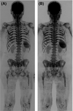
Figure 8.

False positive whole body diffusion weighted magnetic resonance imaging (WB DW‐MRI) following granulocyte colony‐stimulating factor (GCSF) administration. b900 WB DW‐MRI Maximum Intensity Projection images in a 67‐year‐old female with relapsed multiple myeloma (A) show diffuse abnormal marrow signal indicating infiltration which was confirmed by 20% plasma cell infiltration on trephine. b900 WB DW‐MRI Maximum Intensity Projection images 6 months later following treatment (B) show stable appearances; however; trephine showed regenerating marrow and no plasma cells. The patients had received GCSF 3 d prior to the MRI (B), which caused a false positive MRI secondary to marrow hypercellularity.
