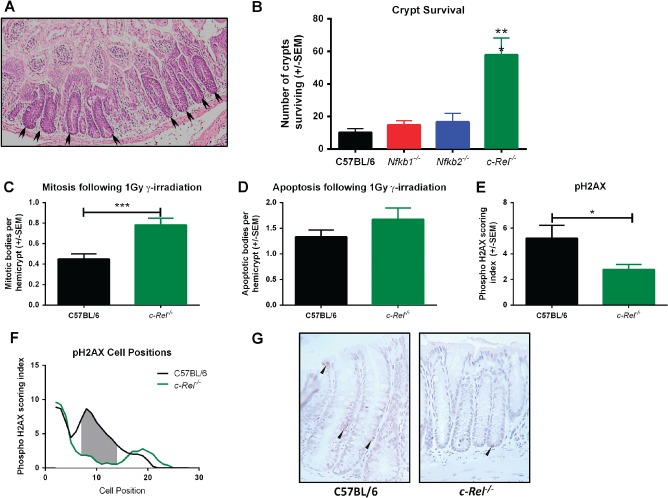
Figure 6.

Crypt survival following γ‐irradiation in the colons of C57BL/6, Nfkb1−/−, Nfkb2−/−, and c‐Rel−/− mice. (A) H&E‐stained sections of c‐Rel−/−colon 96 h following 12 Gy γ‐irradiation. Arrowheads highlight regenerating crypts. (B) Mean number of surviving crypts per circumference of colon 96 h following 12 Gy γ‐irradiation. Differences tested by one‐way ANOVA and Dunnett's multiple comparison test. ***p < 0.001 versus WT (six mice per group). (C–E) Mean percentages of colonocytes from C57BL/6 or c‐Rel−/− mice with morphologically mitotic (C), morphologically apoptotic (D), or expressing pH2AX (E) 4.5 h following 1 Gy γ‐irradiation (two‐tailed Student's t‐test *p < 0.05, ***p < 0.001). (F) Cell positional plot of pH2AX‐expressing cells 4.5 h following 1 Gy γ‐irradiation. Shaded area marks cell positions where a significant difference in pH2AX staining index was detected by modified median test, p < 0.05. (G) Representative photomicrographs of pH2AX staining in colonic mucosa following 1 Gy γ‐irradiation.
