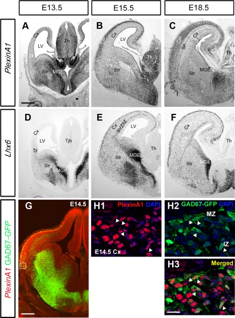
Figure 1.

Expression patterns of PlexinA1 and Lhx6 in wild‐type mouse brain. A–F: In situ hybridization on coronal sections at E13.5 (A,D), E15.5 (B,E), and E18.5 (C,F) for PlexinA1 (A–C) and the interneuron marker Lhx6 (D–F). Overlapping patterns of expression were observed between PlexinA1 and Lhx6 at all ages. G–H3: Coronal section through the brain of a E14.5 GAD67‐GFP mouse, processed for immunohistochemistry with PlexinA1 antibody (red), showed the presence of the receptor within interneurons in the MGE, LGE, MZ, and IZ in the cortex (arrows H1–H3). CP, cortical plate; SP, subplate; Cx, cerebral cortex; IZ, intermediate zone; LV, lateral ventricle; MGE, medial ganglionic eminence; Str, striatum; SVZ, subventricular zone; Th, thalamus. Scale bars = 200 µm in A (applies to A–F); 200 µm in G; 30 µm in H.
