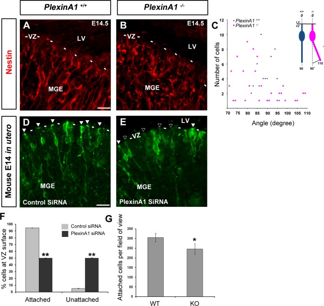
Figure 6.

Reduced attachment and altered morphology of progenitor cells in the MGE of PlexinA1−/− mice. Coronal brain sections from PlexinA1+/+ (A) and PlexinA1−/− (B) mice at E14.5 were immunostained for the progenitor cell marker Nestin (A‐B). (A,B) Fewer Nestin‐positive cells appear to be attached to the ventricular wall and positioned perpendicular to the ventricular surface in PlexinA1−/− mice. (C) Quantification of the orientation of the basal process of labelled cells in the VZ/SVZ in PlexinA1−/− mice. (D,E) Mouse embryos electroporated in utero at E12.5 with control‐(GFP)siRNA (D) or PlexinA1‐(GFP)siRNA (E) into the MGE and harvested at E14.5. Fewer cells appear to be attached to the ventricular surface following PlexinA1 knockdown (black arrowheads) compared to control (white arrowheads), quantified in F. (G) Quantification of adhesion assay, showing fewer E12.5 dissociated MGE cells from PlexinA1−/− mice attached to coated coverslips compared to control littermates. Scale bars in A‐B, 20 μm; and D‐E, 50 μm. (*P < 0.01). Error bars indicate SEM. Abbreviation LV, lateral ventricle; MGE, medial ganglionic eminence; VZ, ventricular zone.
