Figure 7.

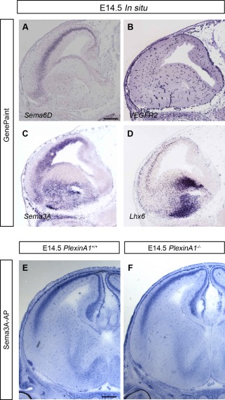
Expression patterns of semaphorins and VEGFR2 in wild‐type mouse brain. A–D: In situ hybridization of parasagittal sections at E14.5 for Sema6D (A) VEGFR2 (B), Sema3A (C), and the interneuron marker Lhx6 (D). Images were downloaded from the GenePaint server. E,F: Sema3A‐AP was added to coronal brain sections from PlexinA1+/+ (E) and PlexinA1−/− (F) mice at E14.5. Similar levels of Sema3A‐AP binding were observed in both sets of animals. Scale bars = 300 µm in A (applies to A–D); 200 µm in E (applies to E,F).
