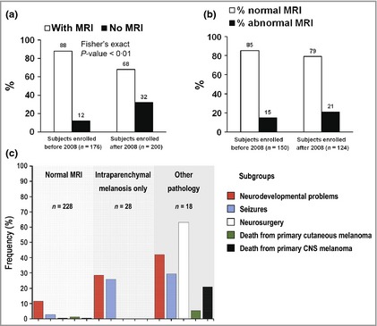
Figure 2.

(a) Comparison of the percentage of patients in whom a magnetic resonance imaging (MRI) scan was performed (white column) and not performed (black column) before and after 2008. By excluding those with only a single congenital melanocytic naevus (CMN), independent of size or site, the introduction of guidelines in 2008 has significantly reduced the percentage of patients scanned routinely. However, the percentage of abnormal scans is not significantly altered, suggesting that we have become more efficient at detecting the same rate of abnormalities. (b) Comparison of the percentage of patients with a normal MRI result (white column) with those with an abnormal result (black column) before and after 2008. The introduction of guidelines in 2008 has not significantly altered the percentage of abnormal scans detected, which implies that we are not failing to detect significant numbers of abnormalities. (c) Subclassification of the radiological abnormalities in this cohort of children with CMN and correlation with the incidence of the different clinical outcome measures in each group. CNS, central nervous system.
