
Figure 1.
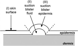
Schematic illustration of collection of skin samples (not shown to scale). Samples from the skin surface (I) were obtained using adhesive sampling discs (D‐Squames®). After raising suction blisters, the blister fluid (II) was collected using a sterile syringe. In the last step, suction blister epidermis (III) was harvested using sterile forceps and scissors (III).