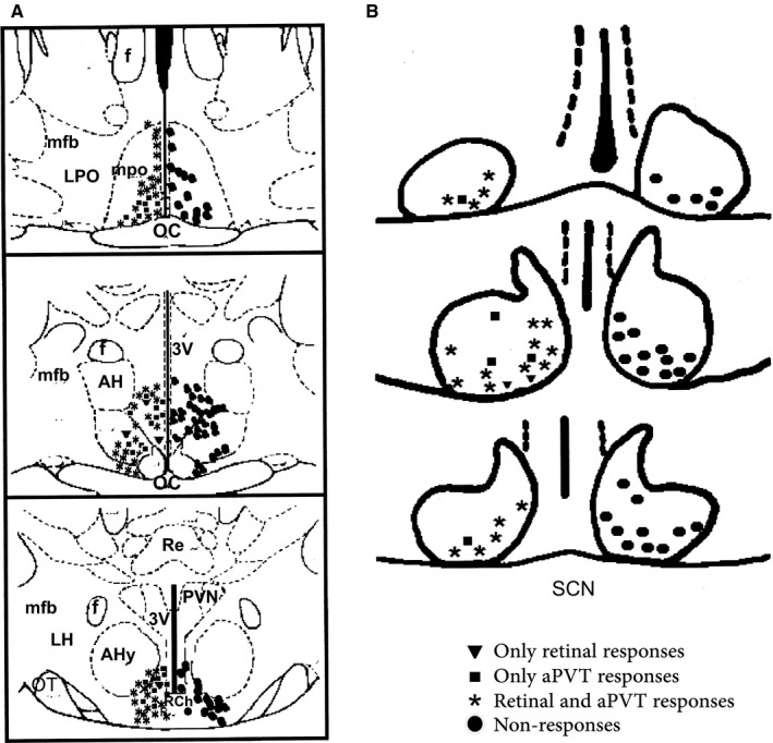
Figure 2.

Location of extracellular recording marks in vivo. (A) Neurons recorded outside the SCN, circles on the right indicate the neurons that did not respond to stimulation; on the left, are neurons that responded to only retinal stimulation, to only aPVT stimulation and to both stimuli. (B) Neurons recorded in the SCN. Abbreviations: OC, optic chiasm; AHy, anterior hypothalamic area; RCh, retrochiasmatic area; mpo, medial preoptic area; LPO, lateral preoptic area; 3V, third ventricle; AH, anterior hypothalamus; F, fornix; PVN, paraventricular nucleus of the hypothalamus; LH, lateral hypothalamus.
