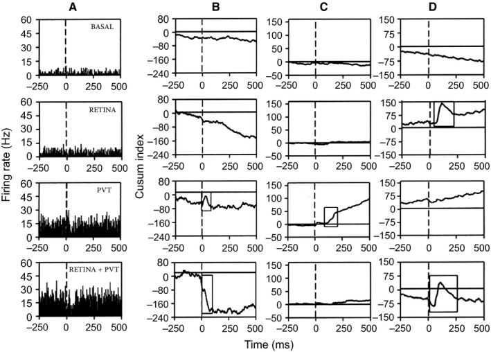
Figure 3.

Examples of 3 SCN neurons recorded in vivo on which functional convergence of the retina and the aPVT occurs. (A) Peristimulus histogram showing a SCN neuron responding to aPVT but not to retinal stimulation; when both afferents were stimulated, the response to aPVT stimulation was modulated from an excitation to an inhibition. (B) CUSUM analysis (cumulative frequency analysis) from the same neurons as in A. (C and D) Two additional examples of the CUSUM analysis; in C, the neuron responded to aPVT stimulation with an excitation that disappears when the aPVT is stimulated simultaneously with the retina. In D, the SCN neuron responded to retinal stimulation with an excitation followed by inhibition; simultaneous stimulation of both the aPVT and the retina modulates the response to inhibition‐excitation‐inhibition. The rectangles in the CUSUM graphs indicate the statistically significant responses (Mann‐Whitney test, P < 0.05).
