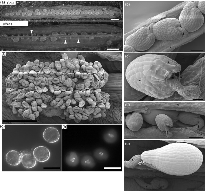
Figure 2.

Ovule abortion phenotype of eif4a1 mutant plants.
(a) Dissected Col‐0 and eif4a1 siliques.
(b–e) Scanning electron micrographs of ovules.
(b) Col‐0 ovules of uniform size.
(c) Pollen tube (arrowed) growing over an eif4a1 mutant ovule.
(d) Three small abnormal ovules overlying a normal ovule in an eif4a1 mutant silique.
(e) An abnormal ovule that has disintegrated and a normally developing ovule in an eif4a1 mutant silique.
(f) A scanning electron micrograph of a dehiscing eif4a1 mutant anther.
(g, h) 4′,6‐Diamidino‐2‐phenylindole (DAPI)‐stained pollen grains seen by Nomarski and epifluorescence microscopy, respectively. Scale bars: (a) 1 mm; (b, d, e, f) 100 μm; (c) 25 μm; (g, h) 15 μm.
