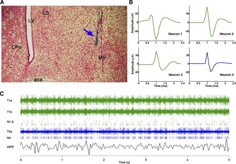
Fig. 1.
Single-unit recording in the medial septum (MS). A: histological reconstruction of the electrode track (blue arrow) in the MS in 1 experiment. aca, Anterior commissure; CPu, caudal putamen; LS, lateral septum; LV, lateral ventricle. B: waveform of 4 simultaneously recorded units in the MS; 3 units (neurons 1–3, green) were separated from 2 wires of tetrode 1 and the 4th (neuron 4, blue) from a single electrode of tetrode 2 (y-axes use the same scale for all units). C: 3 traces of original recording from tetrode 1 (T1a and T1b) and from tetrode 2 (T2a) in the MS, spike trains of 4 separated units (N1-3 and N4), and hippocampus (HIPP) local field potential (LFP) during a 5-s recording.

