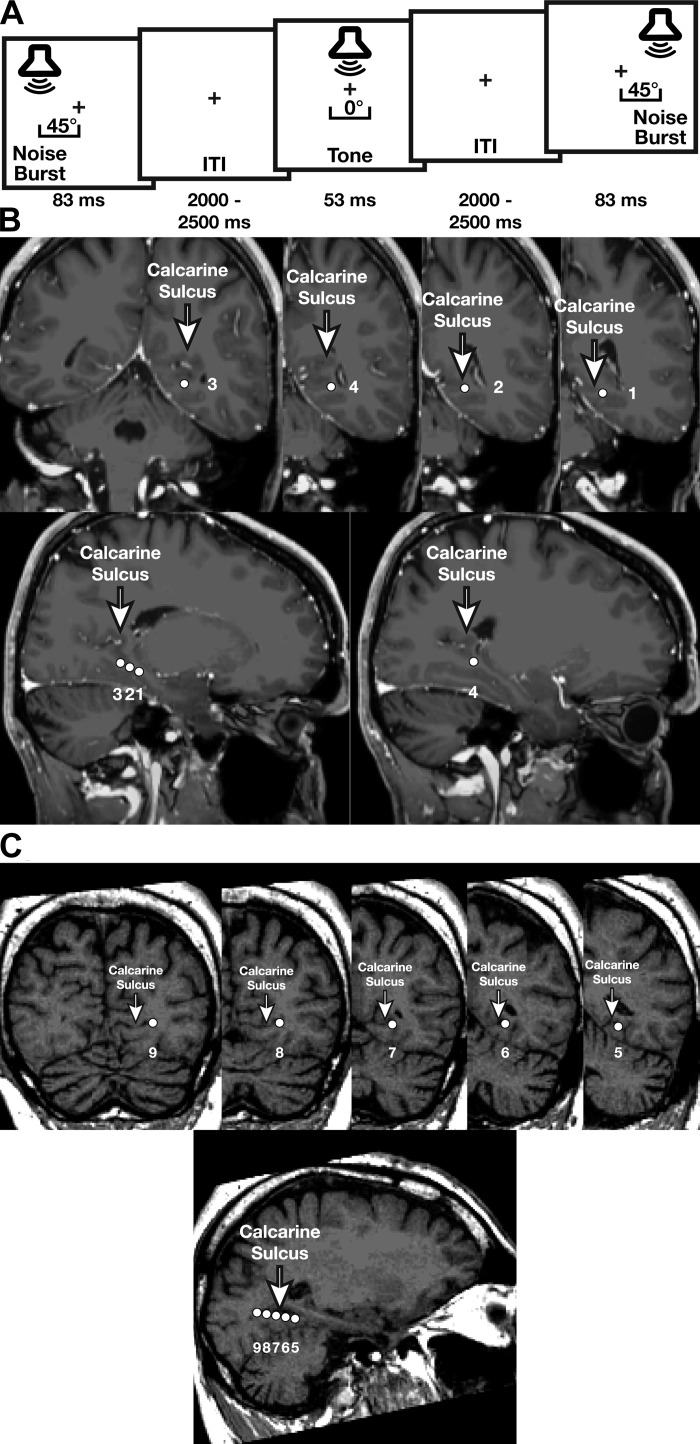
Fig. 1.
A: auditory detection paradigm. Patients responded to a central tone and withheld responses to peripheral noise bursts, allowing measurement of auditory-evoked visual responses to contralateral and ipsilateral sounds without response-related neural activation. ITI, intertone interval. B and C: coronal and sagittal images showing electrode locations (1-9) relative to the calcarine sulcus for patients 1 (B) and 2 (C).

