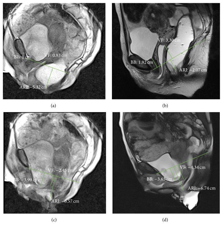
Figure 4.
MR Defecography. Rest phase in sitting (a) and supine (b) position. Evacuation phase in sitting (c) and supine (d) position. The pathological fixed descent was detected only in sitting position in rest phase (a). In evacuation phase, the MR examination in supine position overestimates the dynamic descent; the rectocele is seen only in sitting position. BB: bladder base; VF: vaginal fornix; ARJ: anorectal junction.

