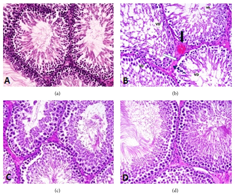
Figure 5.
(a) Photomicrograph of the testis of control rat showing normal structure of seminiferous tubules containing different types of spermatogenic cells (H&E, ×400). (b) Photomicrograph of the testis of rat that received arsenic showing deformed Leydig cells (white arrow), vacuolated spermatogenic cells (VD), thickened basement membrane (dotted arrow), and congestion of blood vessel (black arrow) (H&E, ×400). (c) Photomicrograph of the testis of rat treated with S. platensis + As showing normal spermatogenesis and cell arrangement (H&E, ×400). (d) Photomicrograph of the testis of rat treated with S. platensis showing normal structure (H&E, ×400).

