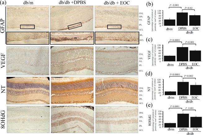
Fig 1. Intravenous injection of EOCs prevented diabetic retinopathy and oxidative retinal damage markers in db/db mice.
(a) Representative photomicrograph of glial reactivity (GFAP), VEGF, nitrotyrosine (NT) and 8-OHdG in retinal tissue from mice treated with EOC. Magnification X400. (b-e) Semiquantitative analyses of immunolabeling.The semi quantitative analyses were performed using the Leica Application Suite (Leica Microsystems, Wetzlar, Germany) in nine non-consecutive retinal sections divided among three slides per animal, per group, under high-powered microscopic viewing (×400) (Zeiss, Jena, Germany).

