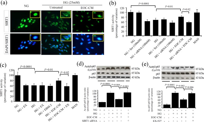
Fig 4. EOC-CM improved SIRT1 deacetylation pathway in rMC-1 exposed to high glucose conditions.
(a) Immunofluorescence for SIRT1 in rMC-1 cultured for 24 hours. In normal glucose (NG), the SIRT1 signal was stronger and present in the nucleus; in high glucose (HG), the staining becomes faint and appears to be located predominantly in the cytosol; magnification X1000. (b-c) SIRT1 activity in rMC-1 cultured for 24 hours under different conditions was estimated by the fluorescent method. (d-e) Representative Western blots for acetyl-Lys310-RelA/p65 in rMCs-1 exposed to HG in presence or not of EOC-CM combined or not with SIRT1 siRNA (d) or EX-527 (e). Equal loading and transfer were ascertained by reprobing the membranes for ß-actin.

