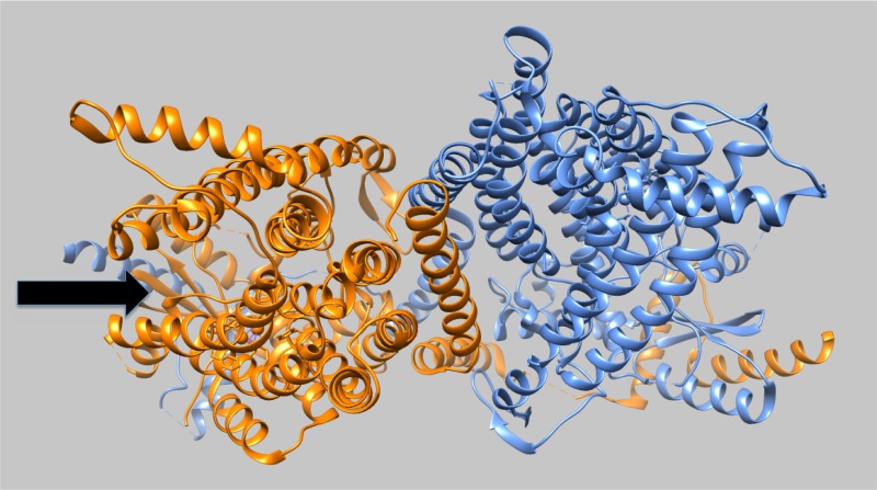
Figure 1.
TMEM16 structure. The structure of the Nectria haematococca TMEM16 dimer is shown as viewed from the membrane outer surface. The symmetry axis is tilted slightly toward the top of this figure to reveal the hydrophilic trench (arrow) on the protein surface that extends through the membrane core and forms the likely path taken by the phospholipid headgroups in this Ca2+-activated form of the scramblase. A similar groove is present in the other subunit but is obscured by the tilting of the viewing axis. Image created from PDB 4WIS35 using Chimera (https://www.cgl.ucsf.edu/chimera/).

