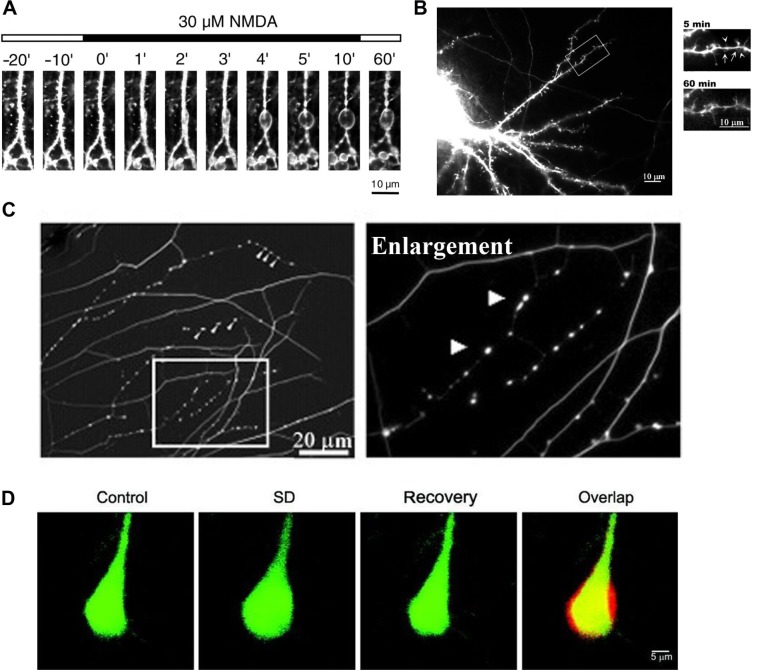
Figure 1.
Formation of neuroprotective endogenous varicosities: (A) confocal laser microscopy images of formation of dendritic varicosities in rat hippocampus neurons treated with 30 μM NMDA;255 (B) immunofluorescent images of formation of dendritic varicosities (arrows) in rat embryo hippocampus neurons, immediately after exposure to 30 μM NMDA (5 minutes) and reversal of varicosities after recovery (60 minutes);14 (C) immunohistochemistry image stained for tubulin, showing varicosity formation in embryonic DRG neurons in response to resiniferatoxin activation of TRPV1;18 (D) two-photon laser scanning images, showing the transient increase in mouse neuron volume before (control), during spreading depression (SD) and after recovery from SD, including a merged image (overlap) showing the overlap (yellow), before volume (green), and during CSD (red).259

