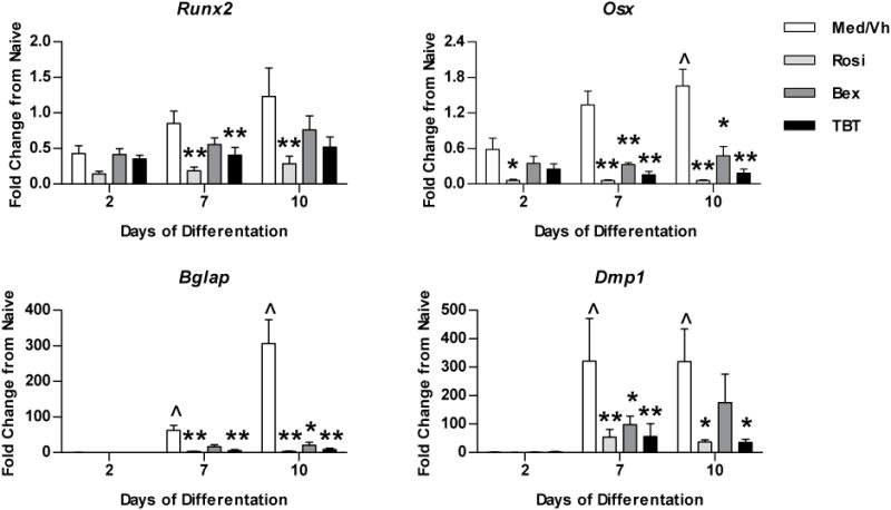
Figure 3. PPARγ and RXR ligands suppress osteogenic gene expression in BM-MSCs.

Primary bone marrow cultures were established and osteogenesis was initiated as in Figure 2. Cells were treated with Vh (DMSO, 0.1%), rosiglitazone (Rosi), bexarotene (Bex) or TBT (100 nM), cultured for 2–10 days and analyzed for gene expression by RT-qPCR. Data are presented as means ± SE (n=4–8). Statistically different from day 2, Vh-treated (ˆp<0.05, ˆˆp<0.01, ANOVA, Dunnett’s). Statistically different from Vh-treated on the same day (*p<0.05, **p<0.01, ANOVA, Dunnett’s).
