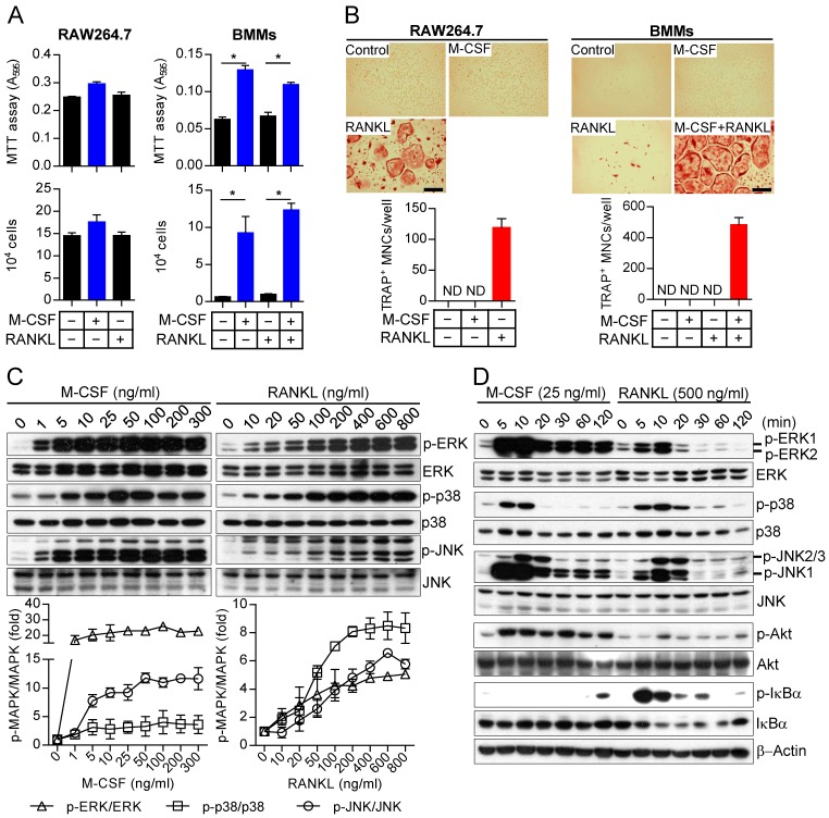
Figure 1.
MAPK activation by M-CSF or RANKL in osteoclast precursors. (A) RAW264.7 cells and BMMs as osteoclast precursors were treated with M-CSF (30 ng/mL) or RANKL (100 ng/mL) as indicated for 2 days, after which cell proliferation was evaluated with the MTT assay or by cell counting. Data between groups were analyzed by one-way ANOVA comparison from a representative experiment run in triplicate. *P < 0.01. (B) RAW264.7 cells and BMMs as osteoclast precursors were cultured in the presence of M-CSF (30 ng/mL) or RANKL (100 ng/mL) as indicated for 4 days, after which the cells were stained for TRAP and the number of TRAP-positive multinucleated cells (TRAP+ MNCs) with more than three nuclei was counted. Data are means ± SD (n = 3) for three independent experiments. ND, none detected. Representative images of the stained cells are also shown. Scale bar, 100 µm. (C) Osteoclast precursors (BMMs) were treated with various concentrations of M-CSF or RANKL for 10 min, after which cell lysates were prepared and subjected to immunoblot analysis with antibodies to total or phosphorylated (p-) forms of the MAPKs ERK, p38, and JNK. The blots were also subjected to densitometric analysis for determination of each p-MAPK/MAPK ratio. The quantitative data are means ± SD (n = 3) of three independent experiments with similar results. Overall P value for fold induction by M-CSF or RANKL stimuli compared to unstimulated control is < 0.05 (Student t test). (D) Osteoclast precursors (BMMs) were treated with M-CSF (25 ng/mL) or RANKL (500 ng/mL) for the indicated times, after which cell lysates were subjected to immunoblot analysis with antibodies to total or phosphorylated forms of MAPKs, Akt, and IκBα as well as to β-actin (loading control). The gel images are representative of three independent experiments.

