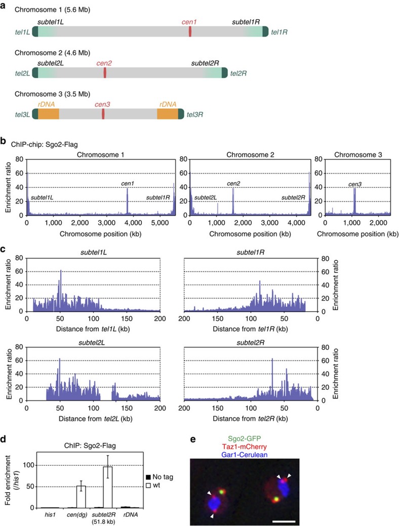
Figure 1. Sgo2 associates with the whole subtelomeres of chromosomes 1 and 2.
(a) Schematic illustration of S. pombe chromosomes. Chromosomes 1 and 2 contain telomere-proximal common sequences that are highly similar among the subtelomeres, whereas chromosome 3 contains rDNA repeats close to the telomeres. (b) ChIP–chip analysis of the genome-wide distribution of Sgo2-Flag in logarithmically growing wt cells. It is noteworthy that the probes used in the tiling array did not encompass regions outside the rDNA repeats on chromosome 3. (c) Magnification of the ChIP–chip data for the subtelomeric regions shown in b. Gaps in the data represent regions not included in the chips due to a lack of sequence information. (d) ChIP analyses of Sgo2-Flag localization at the rDNA repeats in asynchronous cultures (predominantly in interphase). Relative fold enrichment, normalized to the signal at the his1+ locus, is shown. No tag indicates the negative control for the ChIP analyses; cen(dg) indicates the dg repeats at the centromeres; subtel2R-51.8K indicates 51.8 kb from tel2R. Error bars indicate the s.d. (n=3). (e) Subnuclear localization of Sgo2-GFP. Sgo2-GFP, Taz1-mCherry (telomere) and Gar1-Cerulean (nucleolus) signals are shown in green, red and blue, respectively. Arrowheads indicate the telomeres of chromosome 3. Scale bar, 2 μm.

