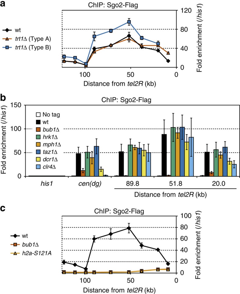
Figure 3. H2A-S121 phosphorylation by Bub1 is important for Sgo2 association with the subtelomeres.
(a) ChIP analyses of Sgo2-Flag localization at subtel2R in trt1Δ survivor cells (Types A and B). Relative fold enrichment at subtel2R, normalized to the signal at the his1+ locus, is shown. It is noteworthy that the most telomere-proximal data point, which corresponds to the region lost in Type B, is missing for Type B. (b) ChIP analyses of Sgo2-Flag localization in various deletion mutants. Relative fold enrichment at subtel2R and the centromeres (dg), normalized to the signal at the his1+ locus, is shown. (c) ChIP analysis of Sgo2-Flag localization at subtel2R in the bub1Δ and h2a-S121A mutants. The relative fold enrichment at subtel2R as normalized by the signals at the his1+ locus is plotted. The error bars indicate the s.d. (n=3).

