Figure 4.
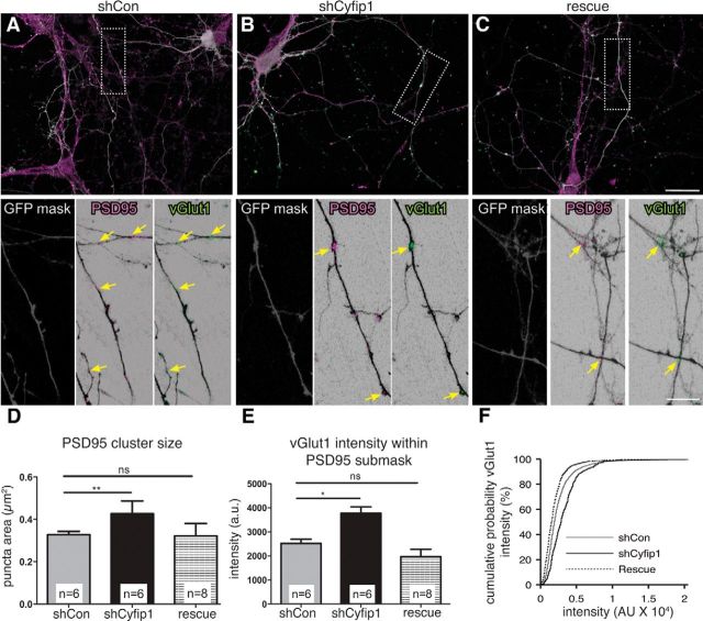
Presynaptic Cyfip1 regulates synapse size. A–C, Confocal images of 10 DIV neurons expressing shCon, shCyfip1, or shCyfip1 + hCyfip1 together (rescue) and immunolabeled as indicated. Low-magnification views represent GFP (white), PSD95 (magenta), and vGlut1 (green) as an overlay. High-magnification views of areas within dotted lines represent a mask of GFP labeling (gray) used to demarcate axons; PSD95 (magenta) and vGLUT1 (green) labeling is shown within the GFP mask. Yellow arrows indicate sites with both labels. Scale bars: overlay, 25 μm; inset, 15 μm. Bar graphs compare sizes of PSD95-immunolabeled clusters within transfected axons (D; ANOVA, p = 0.003) and vGlut1 labeling intensity at PSD95 sites (E, F; ANOVA, p = 0.0006). Differences observed with shCyfip1 are restored to controls conditions by coexpression of hCyfip1 (rescue). *p < 0.05; **p < 0.01. ns, Not significant.

