Figure 6.
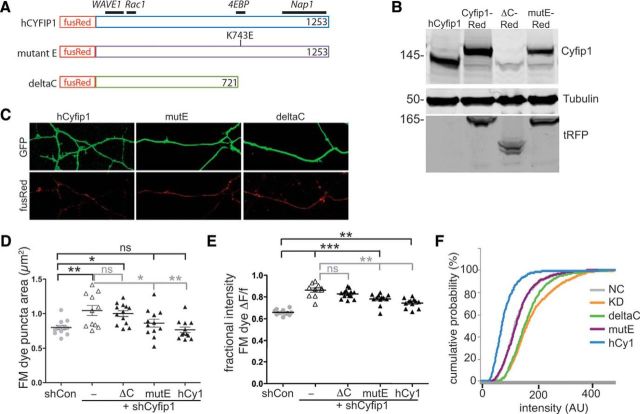
Cyfip1-WAVE1 interaction regulates presynaptic function. A, Maps of relevant Cyfip1 binding partners and the mutants that were generated from human Cyfip1 fused in-frame to a N terminus FusionRed fluorescent tag (Evrogen). B, Western blots of whole-cell lysates generated from HEK cells expressing the cDNAs indicated and blotted for the antibodies shown at right. The antigen recognized by anti-Cyfip1 is compromised in the ΔC mutant, but the truncated Cyfip1 mutant can be detected by an anti-tRFP, which recognizes fusion Red. C, Thirty-six hours following transfection, red fluorescence shows that all expression constructs encode fusion protein and that the mutants are expressed as well as the WT human Cyfip1 in neurons in which shCyfip1 (green) is expressed. D, E, Scatter plots show impact of shCyfip1 knockdown alone and together with the indicated mutant and full-length cDNA constructs in 10 DIV rat hippocampal neurons on FM dye labeling. D, Plot of puncta area following uptake. E, Plots of mean (Fuptake − Frelease)/Fuptake. Horizontal black bars are compared with shCon. Gray bars are compared with shCyfip1 alone. F, A cumulative probability plot of uptake intensity reveals that expression of the WT human Cyfip1 (hCy1) along with shCyfip1 shifts the curve leftward relative to control neurons and underscores similarities between control and mute-expressing neurons. Tukey's post test values, *p < 0.05, **p < 0.01, ***p < 0.001.

