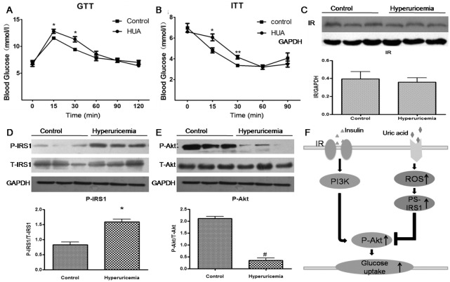
Fig 6.
(A-B) Glucose tolerance test (GTT) (A) and insulin tolerance test (ITT) (B) in an acute hyperuricemic mice model (with HUA level). Data are mean±SD from 3 separate experiments. *P < 0.05, **p < 0.01 vs. control. (C-E) Western blot analysis of IR (C), phospho-IRS1 (Ser307) (D) and phospho-Akt (E) level in cardiac tissues. *P<0.01 vs. control. #P<0.01 vs. control. (F) Schematic representation of HUA-mediated insulin resistance in cardiomyocytes. Increased HUA-induced oxidative stress activates phospho-IRS1 (Ser307) level, which impairs Akt (Ser 437) phosphorylation, thus increasing acute insulin resistance in cardiomyocytes with HUA treatment.

