Figure 1.
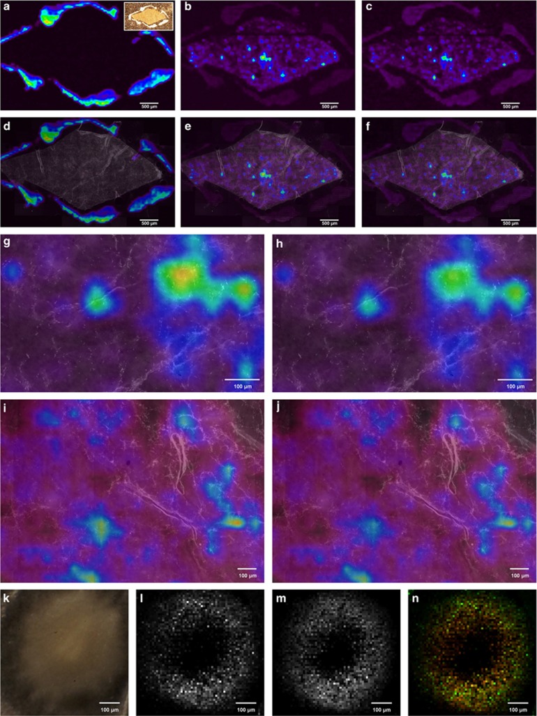
(MA)LDI-FISH of antibiotics produced by symbiotic ‘Streptomyces philanthi' bacteria on a beewolf cocoon (Philanthus triangulum) and in vitro. Ion-intensity maps of (a) the paint marker for alignment of LDI and FISH pictures (m/z 322.5); inset: image of the cocoon piece surrounded by white paint markings on the LDI target plate, (b) piericidin A1 (PA1, m/z 454.5 [M+K]+) and (c) piericidin B1 (PB1, m/z 468.5 [M+K]+). (d–f) The same maps, overlayed with a FISH micrograph of the cocoon piece. Symbiont cells were labeled with the fluorescent oligonucleotide probe SPT177-Cy5. (g–h) Magnifications of (e, f), respectively, with individual bacterial cells visible. (i–j) MALDI-FISH of (i) PA1 (m/z 454.5 [M+K]+) and (j) PB1 (m/z 468.5 [M+K]+) on another cocoon piece. (k–n) AP-SMALDI imaging of antibiotics produced by ‘S. philanthi' in vitro. (k) Light microscopic image of an ‘S. philanthi' colony, (l) PA1 (m/z 416.27 [M+H]+), (m) PB1 (m/z 430.25 [M+H]+), (n) overlay of PA1 (green) and PB1 (red).

