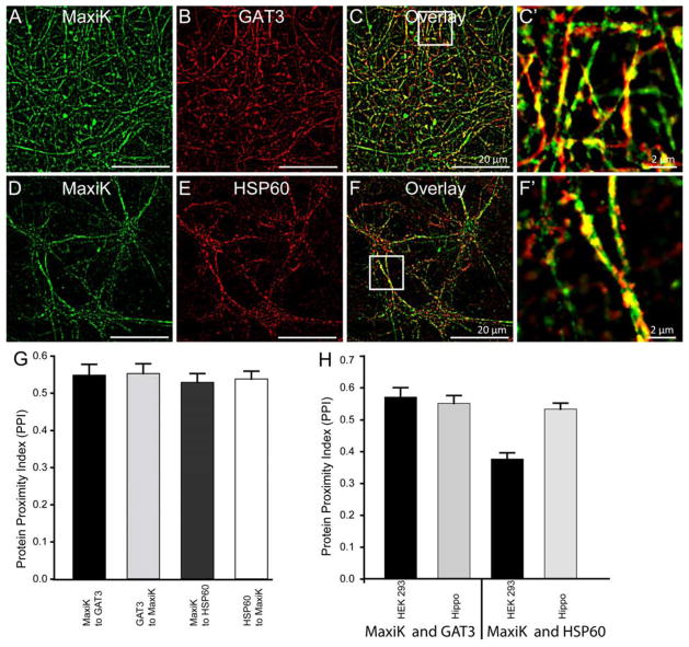
Figure 5. MaxiK channel colocalizes with GAT3 and HSP60 in primary hippocampal neurons.
Primary hippocampal neurons were labeled with anti-MaxiK (A) and anti-GAT3 antibodies (B). C. Overlay image of A and B showed high degree of colocalization of MaxiK channel and GAT3 in neurons. C′ is enlarged squared region from C. Neurons were also colabelled with anti-MaxiK (D) and anti-HSP60 antibodies (E). As seen in HEK293T cells, HSP60 and MaxiK channel also colocalizes in primary neurons (F). F′ is an enlarged squared region from F. G. Bar graph representing colocalization index for MaxiK channel with GAT3 (black), GAT3 with MaxiK channel (light grey), MaxiK channel with HSP60 (dark grey) and HSP60 with MaxiK channel (white) in hippocampal neurons. H. Bar graph representing colocalization between MaxiK channel and GAT3 as well as MaxiK channel and HSP60 in HEK293 and hippocampal neurons, respectively.

