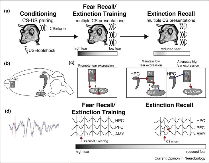
Figure 1.
Behavioral fear paradigms and their anatomical and physiological correlates. (a) Behavioral paradigms for fear learning and memory. Rodents learn to associate a neutral tone (conditioned stimulus, CS) with an aversive outcome, a footshock (unconditioned stimulus, US). Learning for this association is measured by cessation of movement (freezing). Memory for the association is measured at a later time point during which the CS is presented in the absence of the US. During the fear recall trial, the animal expresses high freezing/fear, displaying its memory for the CS–US pairing. As extinction trials (CS exposure without the US) progress, the animal learns that the CS no longer predicts the US, and freezing decreases. Memory for extinction can be tested in a subsequent session by assessing freezing to the CS during an extinction recall session. (b) Structural representation of the areas important in fear learning. The hippocampus (HPC), prefrontal cortex (PFC), and amygdala (AMY) are the main interconnected regions of the fear circuit. The AMY regions depicted include basolateral (BLA), central nucleus (CeA) and the intercalcated cells (ITCs). PFC is divided into prelimbic (PL) and infralimbic (IL) subdivisions. (c) Circuits active during extinction learning and extinction recall. During states of high fear during the fear recall/extinction training session PL activates BLA neurons, leading to excitatory output from CeA and fear expression. Activation of 2 pathways inhibits fear expression during extinction recall. To inhibit fear expression, HPC activates IL, which projects to the GABAergic ITC neurons and inhibits fear output from CeA. To decrease activity of the extinction fear expression circuit, the HPC inhibits PL, leading to an indirect decrease of CeA output. (d) The physiological activity correlated with fear memory and learning. Left, an example of a raw LFP trace with data filtered between 1 and 12 Hz to display the increase in theta activity following freezing behavior. During states of high fear theta frequency activity is synchronized across the fear circuit. During extinction recall, there is less theta synchrony in response to the CS. In addition, theta phase activity in PFC leads the AMY, which is hypothesized to be a signal of learned safety (see text).

